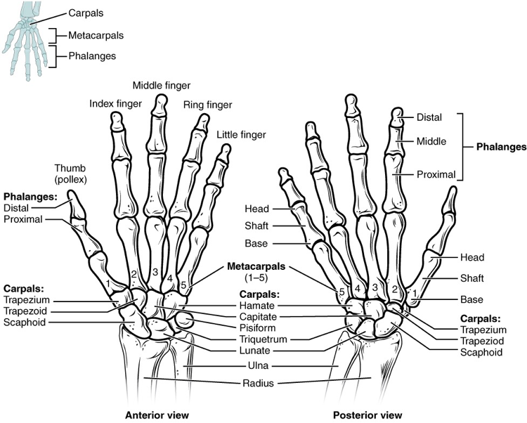
The wrist and base of the hand are formed by a series of eight small carpal bones (Picture 1).
The carpal bones are arranged in two rows, forming a proximal row of four carpal bones and a distal row of four carpal bones.
The bones in the proximal row, running from the lateral (thumb) side to the medial side, are:
- scaphoid (“boat-shaped”),
- lunate (“moon- shaped”),
- triquetrum (“three-cornered”),
- pisiform (“pea-shaped”) bones. The small, rounded pisiform bone articulates with the anterior surface of the triquetrum bone. The pisiform thus projects anteriorly, where it forms the bony bump that can be felt at the medial base of your hand.
The distal bones (lateral to medial) are:
- trapezium (“table”),
- trapezoid (“resembles a table”),
- capitate (“head-shaped”),
- hamate (“hooked bone”) bones. The hamate bone is characterized by a prominent bony extension on its anterior side called the hook of the hamate bone.
A helpful mnemonic for remembering the arrangement of the carpal bones is “So Long To Pinky, Here Comes The Thumb.” This mnemonic starts on the lateral side and names the proximal bones from lateral to medial (scaphoid, lunate, triquetrum, pisiform), then makes a U-turn to name the distal bones from medial to lateral (hamate, capitate, trapezoid, trapezium). Thus, it starts and finishes on the lateral side.
The carpal bones form the base of the hand. This can be seen in the radiograph (X-ray image) of the hand that shows the relationships of the hand bones to the skin creases of the hand (Picture 2).
Within the carpal bones, the four proximal bones are united to each other by ligaments to form a unit. Only three of these bones, the scaphoid, lunate, and triquetrum, contribute to the radiocarpal joint. The scaphoid and lunate bones articulate directly with the distal end of the radius, whereas the triquetrum bone articulates with a fibrocartilaginous pad that spans the radius and styloid process of the ulna. The distal end of the ulna thus does not directly articulate with any of the carpal bones.
The four distal carpal bones are also held together as a group by ligaments. The proximal and distal rows of carpal bones articulate with each other to form the midcarpal joint (Picture 2). Together, the radiocarpal and midcarpal joints are responsible for all movements of the hand at the wrist. The distal carpal bones also articulate with the metacarpal bones of the hand.
In the articulated hand, the carpal bones form a U-shaped grouping. A strong ligament called the flexor retinaculum spans the top of this U-shaped area to maintain this grouping of the carpal bones. The flexor retinaculum is attached laterally to the trapezium and scaphoid bones, and medially to the hamate and pisiform bones. Together, the carpal bones and the flexor retinaculum form a passageway called the carpal tunnel, with the carpal bones forming the walls and floor, and the flexor retinaculum forming the roof of this space (Picture 3). The tendons of nine muscles of the anterior forearm and an important nerve pass through this narrow tunnel to enter the hand. Overuse of the muscle tendons or wrist injury can produce inflammation and swelling within this space. This produces compression of the nerve, resulting in carpal tunnel syndrome, which is characterized by pain or numbness, and muscle weakness in those areas of the hand supplied by this nerve.
Common Characteristics of the Carpal Bones
Each bone (excepting the pisiform) presents six surfaces. Of these the volar or anterior and the dorsal or posterior surfacesare rough, for ligamentous attachment; the dorsal surfaces being the broader, except in the navicular and lunate. The superior or proximal, and inferior or distal surfaces are articular, the superior generally convex, the inferior concave; the medial and lateral surfaces are also articular where they are in contact with contiguous bones, otherwise they are rough and tuberculated. The structure in all is similar, viz., cancellous tissue enclosed in a layer of compact bone.