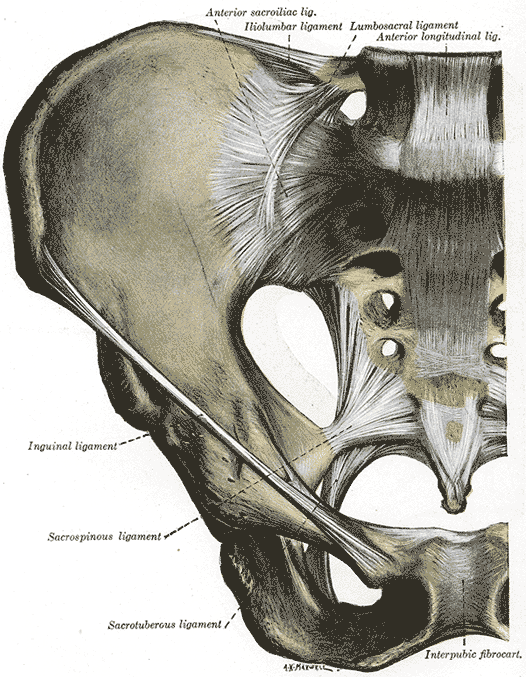
The ligaments connecting the bones of the Pelvis with each other may be divided into four groups and they are around 3 joints:
- [[Sacro-iliac]]:
- Ligaments connecting the sacrum and ilium.
- Ligaments passing between the sacrum and ischium.
- [[SacrococcygealSymphysis]]: with the ligaments uniting the sacrum and coccyx.
- [[PubicSymphysis]]: with the ligaments between the two public bones.
Mechanism of the Pelvis
The pelvic girdle supports and protects the contained viscera and affords surfaces for the attachments of the trunk and lower limb muscles. Its most important mechanical function, however, is to transmit the weight of the trunk and upper limbs to the lower extremities. It may be divided into two arches by a vertical plane passing through the acetabular cavities; the posterior of these arches is the one chiefly concerned in the function of transmitting the weight. Its essential parts are the upper three sacral vertebrae and two strong pillars of bone running from the sacroiliac articulations to the acetabular cavities. For the reception and diffusion of the weight each acetabular cavity is strengthened by two additional bars running toward the pubis and ischium. In order to lessen concussion in rapid changes of distribution of the weight, joints (sacroiliac articulations) are interposed between the sacrum and the iliac bones; an accessory joint (pubic symphysis) exists in the middle of the anterior arch. The sacrum forms the summit of the posterior arch; the weight transmitted falls on it at the lumbosacral articulation and, theoretically, has a component in each of two directions. One component of the force is expended in driving the sacrum downward and backward between the iliac bones, while the other thrusts the upper end of the sacrum downward and forward toward the pelvic cavity. The movements of the sacrum are regulated by its form. Viewed as a whole, it presents the shape of a wedge with its base upward and forward. The first component of the force is therefore acting against the resistance of the wedge, and its tendency to separate the iliac bones is resisted by the sacroiliac and iliolumbar ligaments and by the ligaments of the pubic symphysis. If a series of coronal sections of the sacroiliac joints be made, it will be found possible to divide the articular portion of the sacrum into three segments: anterior, middle, and posterior.
- In the anterior segment (Picture 2), which involves the first sacral vertebra, the articular surfaces show slight sinuosities and are almost parallel to one another; the distance between their dorsal margins is, however, slightly greater than that between their ventral margins. This segment therefore presents a slight wedge shape with the truncated apex downward.
- The middle segment (Picture 3) is a narrow band across the centers of the articulations. Its dorsal width is distinctly greater than its ventral, so that the segment is more definitely wedge-shaped, the truncated apex being again directed downward. Each articular surface presents in the center a marked concavity from above downward, and into this a corresponding convexity of the iliac articular surface fits, forming an interlocking mechanism.
- In the posterior segment (Picture 4) the ventral width is greater than the dorsal, so that the wedge form is the reverse of those of the other segments: the truncated apex is directed upward. The articular surfaces are only slightly concave.
Dislocation downward and forward of the sacrum by the second component of the force applied to it is prevented therefore by the middle segment, which interposes the resistance of its wedge shape and that of the interlocking mechanism on its surfaces; a rotatory movement, however, is produced by which the anterior segment is tilted downward and the posterior upward; the axis of this rotation passes through the dorsal part of the middle segment. The movement of the anterior segment is slightly limited by its wedge form, but chiefly by the posterior and interosseous sacroiliac ligaments; that of the posterior segment is checked to a slight extent by its wedge form, but the chief limiting factors are the sacrotuberous and sacrospinous ligaments. In all these movements the effect of the sacroiliac and iliolumbar ligaments and the ligaments of the symphysis pubis in resisting the separation of the iliac bones must be recognized.
The pelvis during pregnancy
During pregnancy the pelvic joints and ligaments are relaxed, and capable therefore of more extensive movements. When the fetus is being expelled the force is applied to the front of the sacrum. Upward dislocation is again prevented by the interlocking mechanism of the middle segment. As the fetal head passes the anterior segment the latter is carried upward, enlarging the antero-posterior diameter of the pelvic inlet; when the head reaches the posterior segment this also is pressed upward against the resistance of its wedge, the movement only being possible by the laxity of the joints and the stretching of the sacrotuberous and sacrospinous ligaments.